
IJPB / Plant Observatory - Plant Cytology and Imaging Platform
Technological platform
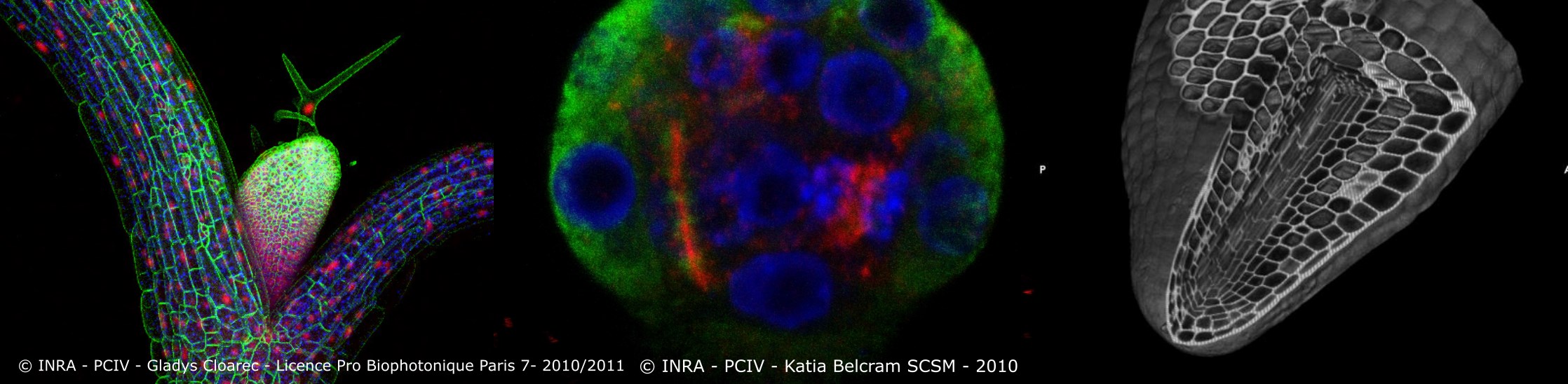
The IJPB's OV-Cytology/Imaging platform brings together a unique range of expertise and instruments dedicated to the cytology and microscopy of plant cells and tissues.
The OV-Cytology/Imaging platform is housed in a 1000 m2 building (B14) and employs 8 permanent staff. It houses all the imaging and cytology activities as well as the equipment needed for the research carried out by the IJPB teams, their partners and external users. It recently welcomed two super-resolution microscopes: a 3D STORM-2 photon-FLIM, the only one of its kind in France, and a Leica Stellaris 8 STED-FLIM.
The OV-Cytology/Imaging platform comprises five workshops:
> Cytogenetics
> In situ hybridization
> Immunocytochemistry
> Microscopy
> Laser microdissection
Each workshop is under the responsibility of permanent specialist staff, who organise the service, welcome and train internal and external users, and prioritise and anticipate technological developments. All the techniques are developed on a large number of plant species, including A. thaliana, Brachypodium, camelina, tomato and maize.
The OV-Cytology/Imaging platform is a leader in France in plant microscopy and cytology. It has pioneered the development of new techniques for studying plants, particularly living cells (e.g. imaging of living meristems, interaction between proteins using fluorescence anisotropy techniques in plants). It is currently developing imaging strategies based on the half-life of fluorescence and techniques for microdissecting different types of tissue in order to carry out spatial transcriptomic analyses. It currently has more than 100 regular users and is a member of the France-BioImaging National Research Infrastructure (https://france-bioimaging.org/about/).
Innovation themes
Lab of attachment
Route de Saint-Cyr
Institut Jean-Pierre Bourgin - Sciences du Végétal (IJPB), Centre INRAE IDF - Versailles-Saclay
78026 VERSAILLES
Technologies utilisées
imaging (brightfield, fluorescence, confocal), cytology, laser microdissection, immunohistology, in situ hybridization, plant imaging, life imagingKeywords
Offres de prestations
Through these activities, the Cyotology and Plant Imaging platform covers a range of techniques, from section to image: traditional cytology, in situ hybridisation, immunolocalisation, cytogenetics and microscopy, brightfield, fluorescence or confocal.
Offres de formations
The platform is integrated into training courses at national level (master's degree and cell biology course of the "Plant Science" doctoral school for UPSaclay and "ABIES" for AgroParisTech) with practical work on confocal microscopes and image analysis stations. Around 400 students and trainees use these facilities every year for visits or practical work in cytology and imaging.
Département(s) de recherche
- SDV (Life sciences)
Liens PIA
- Saclay Plant Sciences LabEx (SPS)
Labellisations
- IBISA